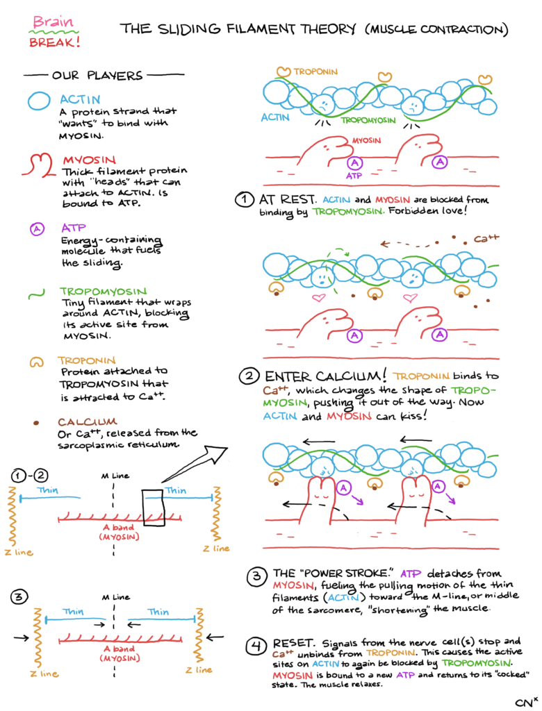
Brain BREAK! presents: The Sliding Filament Theory (Muscle Contraction).
Our players:
- ACTIN. A protein strand that “wants” to bind with myosin.
- MYOSIN. Thick filament protein with “heads” that can attach to actin. Is bound to ATP.
- ATP. Energy-containing molecule that fuels the sliding.
- TROPOMYOSIN. The filament that wraps around actin, blocking its active site from myosin.
- TROPONIN. Protein attached to tropomyosin that is attracted to Ca++.
- CALCIUM. Or Ca++, released from the sarcoplasmic reticulum.
- At rest. Actin and myosin are blocked from binding by tropomyosin. Forbidden love!
- Enter calcium! Troponin binds to Ca++, which changes the shape of tropomyosin, pushing it out of the way. Now actin and myosin can kiss!
- The “power stroke.” ATP detaches from myosin, fueling the pulling motion of the thin filaments (actin) toward the M-line, or middle of the sarcomere, “shortening” the muscle.
* Technically, ATP is in the hydrolyzed form ADP + Pi at this time, but I am simplifying things. - Reset. Signals from the nerve cell(s) stop and Ca++ unbinds from troponin. This causes the active sites on actin to again be blocked by tropomyosin. Myosin is bound to a new ATP and returns to its “cocked” state. The muscle relaxes.
* ATP binds to the myosin, at which point it lets go of actin. Then, as ATP is split into ADP + Pi, it returns to the “cocked” position. During the next contraction cycle, the release of ADP + Pi fuels the “power stroke” that actually pushes the thin filament. Phew!
I like to tell students about this in the love story format because it might stick better. I think of tropomyosin as a protective older brother to actin, and myosin is down below doing a “What light through yonder window breaks?” sort of thing. O myosin, myosin, wherefore art though, myosin? The funny thing about the love story is that myosin is a speed dater. It binds with actin, and as the muscle is destined to contract further, it slides along to the next actin, and on and on. Actin must be pretty jaded by all of these myosins coming along, smooching for a second, and then immediately eyeing her sister.
Also included in this post is a sketch of the rough anatomy of a sarcomere. The myosin filament positioned in the middle is also referred to as the “A band.” The thin filaments form an “I band,” which are attached to a “Z line” on the ends of the sarcomere unit. The thin filaments, pulled along by the myosin heads, move inward, toward the “M line” (I think M for middle). We put “shorten” in quotes to refer to the muscle contraction — my professor stressed for me that nothing’s actually shortening per se. It’s just the sliding motion that creates the tension on the muscle.
I highly recommend watching animated recreations (like this one) so you can see the movement happen. Still images are nice, but models are better for some physiology concepts.
Happy studying,
-CNx